
Questions on muscle contraction
what is the Roles of tropomyosin in muscle contraction
- Tropomyosin regulates muscle contraction by controlling the access of myosin heads to actin filaments.
- At rest, tropomyosin blocks the binding sites on actin, preventing myosin from attaching and initiating contraction.
- During muscle activation, calcium ions bind to troponin, causing a conformational change that pulls tropomyosin away from the binding sites on actin.
- Tropomyosin helps maintain muscle relaxation and prevents unnecessary or involuntary muscle contractions.
what is the Role of calcium ion in skeletal muscle contraction
- Calcium ions are essential for muscle contraction, as they initiate a series of events that lead to the formation of cross-bridges between actin and myosin filaments.
- Calcium ions are released from the sarcoplasmic reticulum in response to an action potential traveling down the T-tubules of the muscle fiber.
- The binding of calcium ions to the protein troponin causes a conformational change that exposes the binding sites on actin, allowing myosin heads to bind and initiate the cross-bridge cycle.
- When the calcium ions are removed, either by reuptake into the sarcoplasmic reticulum or by diffusion out of the muscle fiber, the cross-bridges are broken and muscle relaxation occurs.
Write the molecular mechanism of muscular contraction. Add a note on Rigor Mortis
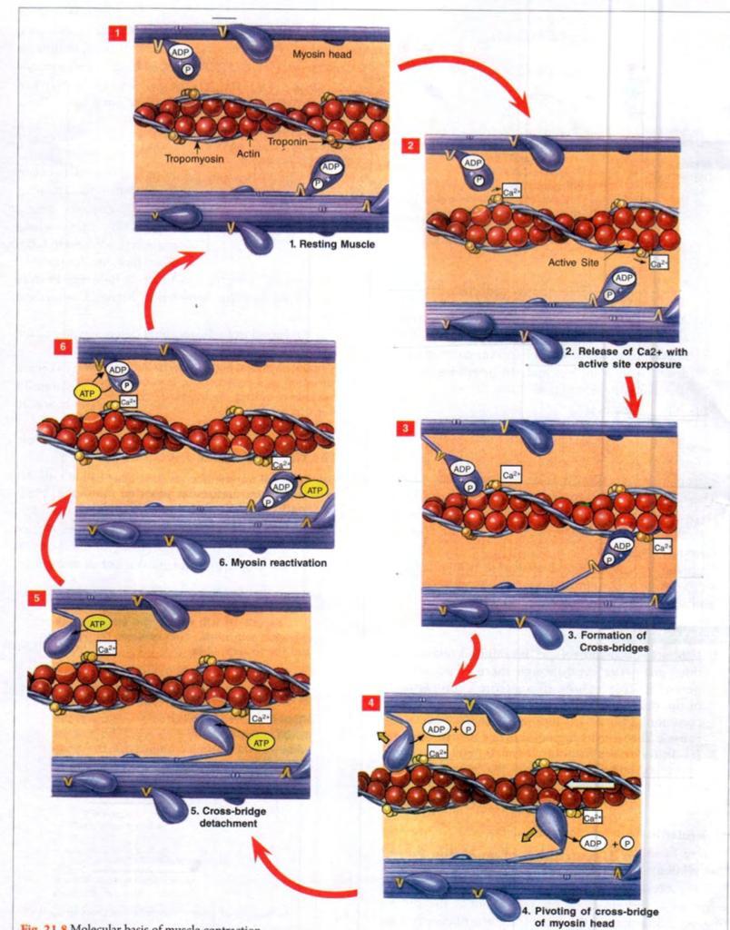
Muscular contraction is a complex process that involves the interaction of various proteins and molecules within the muscle fiber. The molecular mechanism of muscle contraction is best described by the sliding filament theory, which explains how actin and myosin filaments slide past each other to generate force and produce muscle contraction. Here is a detailed explanation of the molecular mechanism of muscular contraction:
Initiation of Muscle Contraction: Muscle contraction is initiated when a nerve impulse reaches the neuromuscular junction, which is the point where the motor neuron connects to the muscle fiber. The nerve impulse triggers the release of acetylcholine, which binds to receptors on the muscle fiber and initiates an action potential that travels along the length of the muscle fiber.
Calcium Ion Release: The action potential reaches the sarcoplasmic reticulum, which is a specialized organelle in the muscle fiber that stores calcium ions. The action potential triggers the release of calcium ions from the sarcoplasmic reticulum into the cytoplasm of the muscle fiber.
Troponin-Tropomyosin Complex: The calcium ions bind to a protein complex known as troponin, which is bound to the protein tropomyosin. The troponin-tropomyosin complex is normally positioned over the binding sites on the actin filament, preventing the myosin head from binding.
Formation of Cross-Bridges: When calcium ions bind to troponin, the troponin-tropomyosin complex undergoes a conformational change that exposes the binding sites on the actin filament. The myosin head then binds to the actin filament, forming a cross-bridge.
Cross-Bridge Cycle: The cross-bridge cycle consists of four stages: attachment, release, power stroke, and reattachment. During the attachment stage, the myosin head binds to the actin filament, forming a cross-bridge. During the release stage, the myosin head releases ADP and Pi, which causes a conformational change that generates force and causes the myosin head to pivot and slide along the actin filament. This is known as the power stroke. During the reattachment stage, the myosin head binds to a new binding site on actin, forming a new cross-bridge. This cycle is repeated multiple times, resulting in the sliding of actin and myosin filaments past each other, which shortens the sarcomere and produces muscle contraction.
Relaxation of Muscle: The process of muscle relaxation is initiated by the removal of calcium ions from the sarcoplasm, which causes the troponin-tropomyosin complex to return to its resting state and block the binding sites on actin. This prevents myosin from binding to actin and initiates the release of cross-bridges, resulting in muscle relaxation
Note on rigor mortis-
Rigor mortis is the stiffening of muscles that occurs after death. It is caused by the depletion of ATP, which is necessary for muscle relaxation. Without ATP, the myosin heads remain bound to the actin filaments, causing the muscles to remain contracted. Rigor mortis typically sets in a few hours after death and can last for several hours or days, depending on the ambient temperature and other factors. Eventually, the muscle tissue breaks down and the body returns to a relaxed state. Rigor mortis is a useful indicator for forensic scientists in determining the time of death.
Name the muscle proteins. What is the role of Troponin C in muscle contraction
There are several types of muscle proteins, including:
- Actin: A thin filament protein that interacts with myosin to produce muscle contraction.
- Myosin: A thick filament protein that interacts with actin to produce muscle contraction.
- Troponin: A regulatory protein that controls the interaction between actin and myosin.
Troponin C plays a crucial role in muscle contraction by binding to calcium ions and initiating a series of events that lead to the movement of myosin along actin filaments.
differentiate between Isotonic and isometric contraction
isotonic contraction
- Muscle length changes during contraction
- Muscle tension remains constant
- Can be divided into concentric and eccentric contractions
- Examples include lifting a weight or performing a bicep curl
isometric contraction
- Muscle length remains the same during contraction
- Muscle tension increases without movement
- No external work is performed
- Examples include holding a weight in a fixed position or pushing against an immovable object
Difference between three types of muscles(skeletal, cardiac and smooth)
Skeletal Muscle
- Also known as striated or voluntary muscle
- Attached to the bones and responsible for the movement of the skeleton
- Under voluntary control
- Cells are long, cylindrical, and multinucleated
- Contraction is rapid and forceful
- Fatigue easily
Cardiac Muscle
- Only found in the heart
- Involuntary muscle
- Cells are branched and connected to each other by intercalated discs
- Cells have a single nucleus
- Contraction is rhythmic and sustained
- Resistant to fatigue
- Can generate its own electrical impulses without nervous system input
Smooth Muscle
- Found in the walls of organs such as the stomach, intestines, and blood vessels
- Involuntary muscle
- Cells are spindle-shaped and have a single nucleus
- Contraction is slow and sustained
- Resistant to fatigue
- Controlled by the autonomic nervous system and hormones
molecular Mechanism of muscle contraction (sliding filament theory)
#MOLECULAR MECHANISM
EVENTS DURING MUSCULAR CONTRACTION
• Electrical events
• Mechanical events
• Chemical events
• Thermal events
ELECTRICAL EVENTS
• Transmission of nerve impulse to muscle across NMJ
• Excitation of muscle→development of muscle action potential
• Transmission of muscle action potential through “T” tubules to interior of muscle fiber
• release of calcium into the cytosol
MECHANICAL OR MOLECULAR EVENTS
• Explained by sliding filament theory-proposed by Huxley and Huxley in 1969. Most accepted theory
• States that muscular contraction is due to sliding of actin filaments over the myosin filaments.
• Involves two important sequences:
- Exposure of myosin binding sites of actin molecules
- Cross bridge cycle
Exposure of myosin binding sites of actin molecules
- • Binding of calcium to troponin C
- • Conformational change of troponin C.
- • Lateral movement of tropomyosin – troponin complex from actin filaments
- • Exposure of myosin binding sites of actin molecules
1. Activation of myosin ATPase enzyme
2. Hydrolysis of an ATP molecule→release of energy→activation of myosin crossbridges
3. Binding of crossbridges to the actin filaments
4. Bending of myosin head when ADP and P are released from the crossbridges → pulls the
actin filament towards the centre of sarcomere (powerstroke)
5. Binding of another ATP molecule→detachment of crossbridges from actin filaments
6. Binding of another ATP molecule → Attachment of crossbridge to another distal actin
molecule
CROSSBRIDGE CYCLE
Attachment – Swivel (power stroke) – detachment – attachment again.
RELAXATION OF MUSCLE
• reuptake of Ca2+ into sarcoplasmic reticulum (SR)
• Troponin-tropomyosin move back to their position and cover the active sites of actin
filaments.
Interaction between the actin & myosin ceases
The muscle relaxes
CHEMICAL EVENTS
• Energy is provided by hydrolysis of ATP & creatine phosphate
Contraction-Relaxation Steps Requiring ATP
• For binding of myosin crossbridges to actin filaments
• Detachment of crossbridges from actin filaments
• Active transport (Pumping) of Ca2+ back into sarcoplasmic reticulum
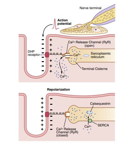
Excitation - contraction coupling
Sequence of Events
The excitation of the nerve leads to excitation of the muscle.
This in turn causes contraction of the muscle. This is referred to
as excitation-contraction coupling. The following steps explain both E-C coupling and mechanism of muscular contraction.
.1 Excitation of the nerve.
.2 Arrival of impulse at the nerve terminal.
.3 Vesicles move and attach to the membrane of sole foot.
4. Release of ACh from the nerve terminal.
5. ACh combines with receptors present on the motor end plate.
6. End plate potential develops.
7. When the end plate potential reaches the firing level, an AP appears on the sarcolemma.
8. Transmission of impulse by transverse tubules into the interior of the muscle fibres.
9. Arrival of impulse at the triads.
10. Voltage-sensitive dihydropyridine protein opens calcium channel (Ryanodine receptor) present in the cisternae.
11. Release of Ca++ from cysternae and Ca++ concentration around thin filaments increases by more than10^-7 moles/L.
12. Ca++ combines with Troponin C
13. Interaction between actin and myosin takes place.
14. Actin slides over myosin causing contractions of the muscles

Describe the Sarcotubular system?
1. It is a highly specialized system of internal conduction of depolarization within the muscle fiber. It is made up of T-system (transverse tubular system) and a longitudinal sarcoplasmic reticulum.
(i) T-System or Transverse Tubular System
(a) They are inwardly directed of the sarcolemma into the muscle fibers extensions at the junction between ‘A’ and ‘T’ bands (A-I junction), therefore, each sarcomere has 2 tubules. Their lumina (30 nm wide) are thus in continuity with the ECF which surrounds the muscle fibrils. Its function is the rapid transmission of the action potential from the cell membrane to all the fibrils in the muscle at the same time.
(b) Depolarization of the T-tubule membrane activates the longitudinal sacroplasmic reticulum (see to below) via dihydropyridine receptors. These receptors are voltage gated Ca2+ channels in the T-tubule membrane and are so called because they get blocked by the drug dihydropyridine. These receptors serve as voltage sensor and cause release of Ca2+ from the longitudinal sarcoplasmic reticulum.
(ii) Longitudinal Sarcoplasmic Reticulum
(a) Longitudinally on either side of the tubular system are found the vesicle or sacs of the dilated longitudinal sarcoplasmic reticulum. These sacs are named terminal cisternae, which are rich in glycogen and Ca2+. It is concerned with Ca2+ movements and muscle metabolism.
(b) The Ca2+ channels in the longitudinal sarcoplasmic reticulum are not voltage gated and allow the release of Ca2+ into the cell. These are called ryanodine receptors, as they are kept in the open position by ryanodine, a plant alkaloid.
2. There is a close proximity i.e. contiguity (and not the continuity) of the terminal cisternae and the T-system. Transverse tubule with two terminal cisterns is called the triad structure which is found at the A-I junction and hence there are two triads per sarcomere. Triad structures are most extensive in muscles which contract and relax very rapidly.